



Our Science
The Next Signal in Skincare
Our exosomes have been imaged, measured and tested on skin. Here is what the research shows.
01The Source
Where our exosomes begin
Our exosomes begin with umbilical cord lining stem cells (ULSCs), a mesenchymal stem cell source selected for its cellular characteristics and secretory profile. In our ingredient partner’s research, these cells formed colonies at a higher frequency than comparator mesenchymal sources and maintained longer telomeres through multiple passages.
The quality of the source cells shapes what they naturally secrete, so every step from donor to finished ingredient is documented and tested.
The donor and the cell line
Single-donor cell line
Cell line 208
Every lot traces back to one fully characterized donor cell line, so the source stays consistent from batch to batch.
Cell identity
> 98% MSC markers
CD90, CD73 and CD44 present in more than 98% of cells; blood-cell markers CD34, CD45 and HLA-DR below 0.3%.
Viral screening
Not detected
PCR panel for human viruses, including hepatitis A, B and C, CMV, EBV and HPV.
Sterility & mycoplasma
Passed
Tested for bacterial and fungal contamination and for mycoplasma.
Endotoxin
< 0.05 EU/mL
Bacterial endotoxin measured in the source material.
Genetic stability
Normal karyotype
Chromosomes analyzed across multiple passages of the cell line.
Cell-free ingredient
No living cells
Carries what the cells naturally secrete, never the cells themselves.
Lot release
GTP / GMP
Every lot is quality-control tested and quality-assurance reviewed before release.
ExoCM® is developed by Exora™ Skin Labs, building on more than 10 years of mesenchymal stem cell research led by Rafael Gonzalez, PhD. Source: Certificate of Analysis, cell line 208 (December 2025).
02The Matrix
More than exosomes
“Exosomes are part of the story. ExoCM® is the matrix around the story.”
Our exosomes are not a suspension of isolated vesicles. They arrive in ExoCM®, a naturally complex secretome: extracellular vesicles together with naturally occurring Collagen I and III, high-molecular-weight hyaluronic acid, beta-defensins, glutathione, peptides and other secreted components.
Inside the matrix
Exosomes
4.96 × 10¹⁰ particles/mL
Nanoscale extracellular messengers that carry proteins, lipids and other signaling cargo.
Collagen I & III
713 / 377 ng/mL
Naturally present structural proteins associated with the appearance of supported, resilient skin.
Hyaluronic acid
1.32 µg/mL · HMW
A naturally occurring moisture-binding component associated with hydration and a supple skin appearance.
Peptides & factors
FGF-2 · LL-37 · IL-1Ra
Characterized secreted factors, including FGF-2 at 71.2 pg/mL and LL-37 at 2.19 ng/mL.
Measured by nanoparticle tracking analysis and protein analysis. Data on file.
03The Signal
Skin speaks in signals
Your skin cells are in constant conversation. Exosomes are one of the ways they communicate: nanometer-sized messengers that carry proteins and growth factors from one cell to the next.
Over time, the cells that build skin’s support structure produce less collagen and elastin. Our exosomes are designed to support that conversation earlier, through biology, not force.
04Seen
Seen. Measured. Characterized.
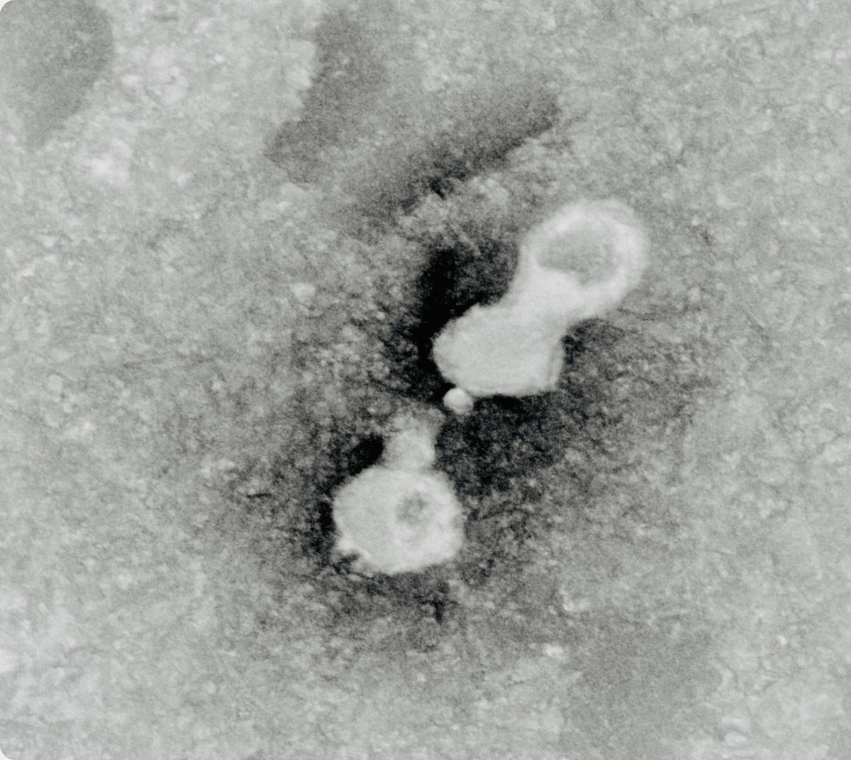
Exosomes are far too small to see with the eye. Ours have been imaged one by one under electron microscopy, and characterized in line with the MISEV guidelines from the International Society for Extracellular Vesicles (ISEV).
Pictured: an electron micrograph of our exosomes.
Measured
9 of 9
ISEV characterization areas reported
81 nm
Typical exosome size
50B
Exosome particles per mL
4
Identity markers confirmed: CD9, CD63, CD81 and TSG101
Measured by nanoparticle tracking analysis, flow cytometry and electron microscopy. Data on file.
05In the Lab
Studied on skin cells
In laboratory testing, skin fibroblasts from a younger (25-year-old) and a mature (53-year-old) donor were exposed to ExoCM® at two concentrations.
Cells from both donors showed measurable, dose-dependent responses associated with collagen and elastin support.
In vitro study on human skin cells. Laboratory results are not a clinical study of a finished product.
Collagen I · secreted
Elastin · secreted
Concentration of our exosomes in culture medium. n.d. = not detected. Redrawn from supplier study data.
06On Skin
Tested on skin, including sensitive skin
Our exosomes were evaluated in a 107-person repeated insult patch test (RIPT), the standard method for assessing irritation and allergic sensitization. Across repeated applications, no reactions were observed. Results were reviewed by a board‑certified dermatologist.
107
Participants completed the study
20–79
Age range of participants
I–VI
All six Fitzpatrick skin types
0
Reactions observed
Two study panels (2025): participants with self-perceived sensitive skin and participants with non-defined skin types.
16
Weeks of stability testing
07Stability
Built to stay intact
Exosomes are delicate. In 16-week stability testing, the proteins carried by our exosomes remained intact, whether refrigerated or kept at room temperature.
Our standard
Essence over excess
Every ingredient is selected for a defined function within the formula. Formulated without fragrance and essential oils, parabens and phthalates, silicones, PEGs, formaldehyde-releasing preservatives and drying alcohols.
References
- Varani J. et al. Decreased collagen production in chronologically aged skin. Am J Pathol. 2006;168(6):1861–1868.
- Zhang S., Duan E. Fighting against skin aging: the way from bench to bedside. Cell Transplant. 2018;27(5):729–738.
- Welsh J.A. et al. Minimal information for studies of extracellular vesicles (MISEV2023). J Extracell Vesicles. 2024;13:e12404.
- Papakonstantinou E. et al. Hyaluronic acid: a key molecule in skin aging. Dermatoendocrinol. 2012;4(3):253–258.
- Data on file: exosome characterization, in vitro fibroblast study, repeated insult patch test (2025) and 16-week stability study.
Studies were conducted on the exosome ingredient used in EUCLASÉ formulations. Individual results may vary.
Say less. Signal more.
Experience our exosomes in the EUCLASÉ Exosome Face Serum.
